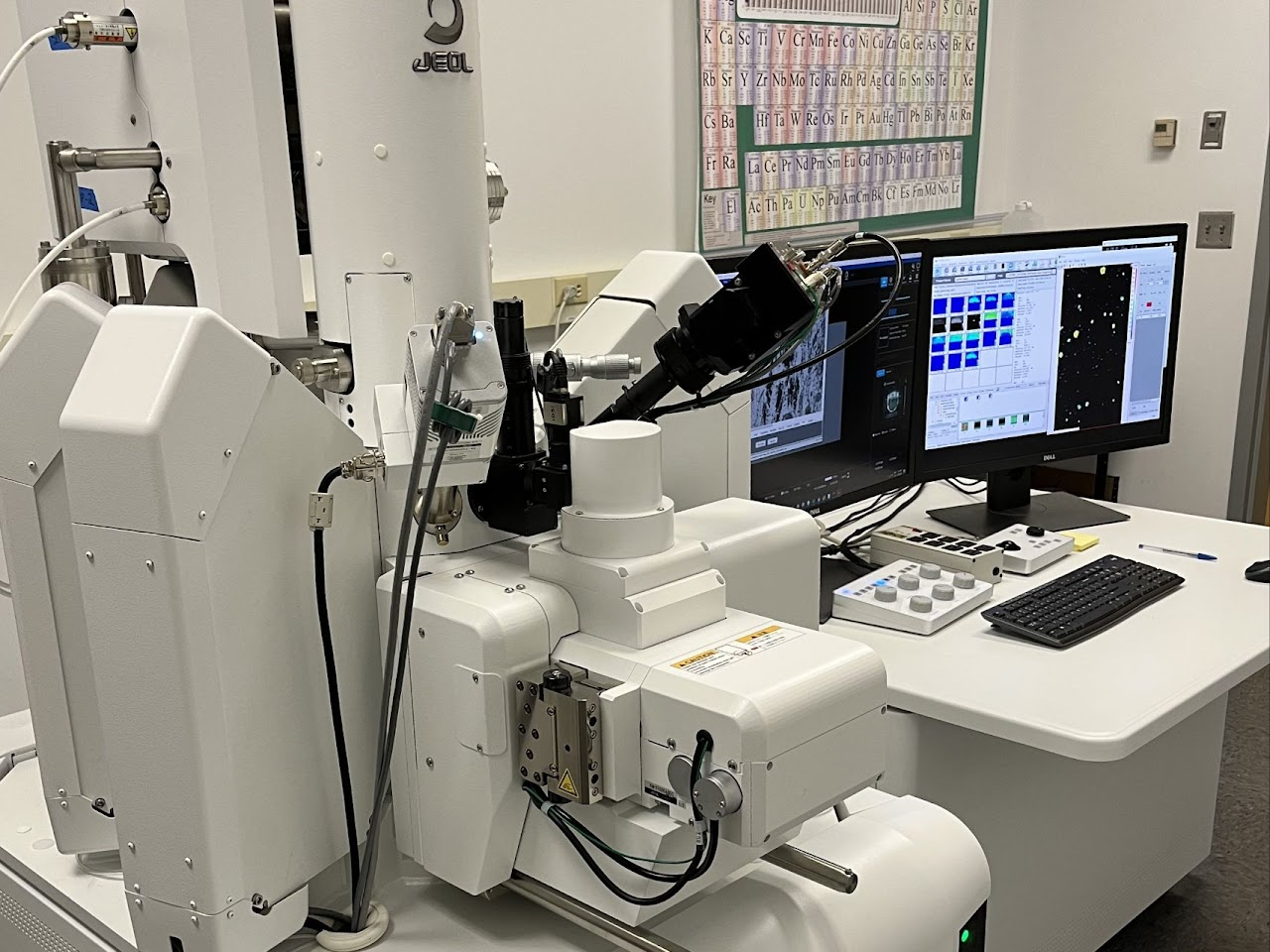
JEOL JXA-iHP200F Field Emission Electron Probe Microanalyzer
To reserve microprobe time, please refer to the scheduling calendar below. Any date not marked as "Reserved" is available. To make a reservation, please email the lab manager (moorelr@vt.edu). Please note that reservations are done by the day (not by the hour), but this does not mean that you have to use or pay for a full day's worth of instrument time.
Note: This calendar sometimes doesn't load correctly, and it appears to depend on the viewer's choice of browser and/or account status with Google (e.g. VT users may need to log out of VT-affiliated Gmail accounts). Here is an alternative public URL for the calendar. A backup static HTML version of the calendar can be found at this URL.
The JXA-iHP200F is located at:
NCFL 1020
Nanoscale Characterization and Fabrication Laboratory
Virginia Tech, Mail Stop 0905
1991 Kraft Drive
Blacksburg, VA 24061
5067 Derring Hall
926 West Campus Drive
Virginia Tech
Blacksburg, VA 24061
Lowell's contact information:
Lowell Moore
Electron Microprobe Lab Manager
Department of Geosciences, Virginia Tech
926 West Campus Dr.
Blacksburg, VA 24061, USA
email: moorelr@vt.edu
office: 5042 Derring Hall
The JEOL JXA-iHP200F field emission electron probe microanalyzer is a state-of-the art tool for chemical mapping and point analysis of materials from the nanometer to millimeter scale. Our new instrument, currently one of two in the United States, has the following features:
Electron probe microanalysis (EPMA) is a conventional term for quantitative chemical analysis performed using wavelength-dispersive x-ray spectroscopy (WDS) with a scanning electron microscope (SEM); a SEM equipped with multiple WDS detectors and specialized electron optics to accommodate them may also be called an electron microprobe (EMP) -- or just a “microprobe."
The WDS detectors involved in EPMA are used to record the spectrum of secondary x-rays produced by a sample being bombarded by a focused electron beam. Unlike detectors used in energy-dispersive spectroscopy (EDS, which may be used to identify X-rays according to their energy), WDS detectors allow characteristic X-rays to be identified based on their wavelength using the principle of X-ray diffraction described by Bragg’s Law. WDS detectors accomplish this by physically moving a crystal with a known atomic unit cell (d-spacing) along a circular path so that only X-rays which satisfy the Bragg equation (nλ = 2dsinθ) are detected. This approach provides superior spectral resolution compared to EDS.
Generally, the microprobe can be used to analyze most solid, inorganic material. Samples must be small enough to fit onto a 2.5 cm wide microscope slide or 1-inch diameter sample holder with a vertical profile not exceeding 1 mm. Unlike a visible light microscope, the analyte observed by the microprobe must be loaded into a vacuum chamber to prevent air from interacting with the electron beam. Additionally, the analyte must remain solid and stable when irradiated by the electron beam (15 kV, 5 nA), which precludes analysis of most organic matter.